Saturday, April 30, 2011

Objective 9: Compare and contrast bond types

I didn't realize how rusty I was on bond types until I reached this objective. I believe I learned this information awhile back when I was in middle school during physical science, came across it again during chemisty, and then brushed by it in anatomy and physiology 1. So, long story short, I had to refer to chapter 2 in our textbook to become refreshed with this old topic. The textbook does a good job of simply explaining what each bond is on pages 27, 28 and 30. There are three major types of chemical bonds: ionic, covalent, and hydrogen bonds.

Ionic bond:

An ionic bond is a chemical bond between atoms where one or more electrons is transferred from one atom to another. The atom that gains one or more electrons is the electron adapter and it obtains a negative charge (anion). The atom that loses one or more electrons is the electron donor and it obtains a positive charge (cation).

Covalent bond:

A covalent bond occurs when two atoms share electrons and the shared electrons occupy a single orbital common to both atoms. When two atoms only share one pair of electrons, a single covalent bond is formed. When two atoms share two or three electron pairs, a double or triple covalent bond is formed.

Hydrogen bond:

A hydrogen bond is more like an attraction than a true bond. Hydrogen bonds form when a hydrogen atom, already covalently linked to one electronegative atom, is attracted by another electron- hungry atom (greedy-grabby) and a "bridge" is formed between them.


These two photos from our textbook is are examples of ionic and covalent bonds.


(ionic bond)


(covalent bond)


While I was surfing the web, I came across this Youtube video describing ionic and covalent bonds and I just had to add it to this objective. It not only is informative, but it is a very cute video that really made this information stick. I hope you enjoy it as much as I did. When I started viewing it, it made me laugh. But be careful, its quite catchy!


Objective 10: Identify classes of hormones

I really thought that this objective would be a piece of cake, but when I tried looking up the classes of hormones online I got a bunch of different answers. So I decided to refer back to the old trusted textbook and chapter 15 lecture outline to find my answer. Hormones are classified into two main classes, which are amino acid-based hormones and steriod hormones. This information is like common sense now after reviewing the slides and textbook, and it makes me laugh that I tried to make this objective harder than it really was. Both classes of hormones are chemical substances that regulate the metabolic function of other cells in the body.


Objective 60 & 61: Pancreatic juices and chemical forms

Objective 60: Describe enzymatic and bicarbonate content of pancreatic juices
Objective 61: State the chemical forms in which the major food classes are absorbed

Reading up on these two topics was a lot easier than I had originally thought. For some reason, I thought that the pancreatic juices were going to be this complicated mix of enzymes and electrolytes. I also believed that the chemical forms of the food classes were going to have a very long process to being absorbed. Well, I was in for a pleasant shock. I was able to located the information in the text book and lecture outlines, and they both did a great job in helping me understand the two. Pancreatic juice consists primarily of water, electrolytes (mainly bicarbonate), and enzymes (amylase, lipases, nucleases). The acinar cells produce the enzyme-rich component of pancreatic juice. Along with the lecture outline slides, I also referred to chapter 22 page 775 to help me understand the make up of the juice.


I copied this chart from chapter 22 lecture outline, but it can also be found on page 785. It really does a great job in outlining the chemical forms that each type of food gets broken down into. It then tells you the path of absorption that these chemicals forms travel within the body. I don't know how I would have figured out this objective without this chart. Its extremely detailed, and informative and it really made that light bulb go off for me. So what I learned is that carbohydrates are broken down into oligosaccharides and disaccharides; protein is broken down into amino acids; fats are broken down into monoglycerides, glycerol and fatty acids; and nucleic acids are broken down into pentose sugars, N-containing bases, and phosphate ions. After the major food classes are broken down into their chemical forms, it is from there that they are absorbed into different parts of the body. The last column of the chart describes in detail where they are shipped off too. Isn't the digestive system spectacular?


Objective 58: Relate structure and function of the endocrine system to digestion

This is a very broad topic, which means there could be a variaty of answers to cover this topic. I actually am still having a hard time figuring out the best way to answer this question correctly, but I'm going to give it my best shot. So, here it goes. The endocrine system is the second great control system of the body. It is made up of a serious of glands that produce a variaty of hormones. These hormones act as "chemical" messengers and help regulate a majority of the body's basic functions. There is a passage from the textbook that really helps me understand, in short, what the endocrine hormones are responsible for. In chapter 15, the passage on page 519 states "The major processes controlled and integrated by these "mighty molecules" are reproduction; growth and development; maintenance of electrolyte, water, and nutrient balance of the blood; regulation of cellular metabolism and energy balance; and mobilization of body defenses." This really helps me see how the endocrine affects the other systems more clearly. The digestion system is composed of a system of organs that helps break down food into nutrients, absorbs the nutrients into the bloodstream, and then gets rid of the wastes. So in short, the endocrine system produces important hormones (for example -gastrin, cholecystokinin, secretin) that allow the digestive function to take place. Without these hormones, our bodies could not digest the food we take in.

I know that we are not suppose to "teach" the information, but I couldn't find anything in the textbook or online that really gave me an "aha" moment. There was a small article called Enteric Endocrine System that provided a few small pieces of information that guided me, but for the most part I was on my own. Being able to write this information out on this blog really gave me a clearer understanding on how the endocrine system was related to the digestion process. The lecture in class was the only helpful thing to help me understand this information, since we discuss certain topics in depth.

Objective 30: Understand how heart valves ensure one-way blood flow during systole and diastole

Where would we be without heart valves? My best guess would be that our heart wouldn't complete systole and diastole correctly, and our heart wouldn't be able to function. Which means a very bad day! The valves in our heart ensure one-way flow of blood, and that is very important to its function! Without these valves the blood flow would back flow into other sections of the heart that they aren't suppose to be in, for example back into the atrium from the ventricles. The textbook was a great reference, and the lecture outline was a great summary of the hearts valves. When I was looking for a good diagram to back-up the textbook's information, I referred to chapter 17 pages 587-588 which provided a diagram of the AV valves and another diagram of the semilunar valves. Each diagram shows the direction of the blood flow into the heart and the blood flow through the valves. They describe how the valves are either forced open or stay closed to make sure that the blood flows one way and does not backflow. These photos were all I needed to understand this concept and I'm glad they were apart of my textbook.


Objective 74: Distinguish between diploid germ cells and haploid sex cells

Understanding mitosis and meiosis has always been an ongoing battle for me. For some reason when I first learned this concept I always got the two mixed up. I found this chart from chapter 26 page 899 that helped me in figuring out the differences between the diploid cell and the haploid cell. Well, I couldn't get a good picture of it from my camera ( I don't have a scanner) so I used the photo from the lecture outlines, but the photo and information is still the same. I promise. This photo also severed a double purpose because it helps me understand mitosis and meiosis. It never hurts to learn extra information!

So, basically the two cells go like this.

Diploid= Double. There are 46 chromosomes in each diploid cell
Haploid=Half. There are 23 chromosomes in each haploid cell

Through fertilization,  two haploid cells combine to produce a zygote. The zygote has now two sets of 23 chromosomes. So 23+23= 46. The zygote now has 46 chromosomes and is a diploid cell. I'm glad that I finally figured out the difference between the diploid and haploid cells. All I seemed to need was a little math.


This video also reinforces the information that the textbook and class lectures gave me about diploid and haploid cells. Still, having an animated diagram allows me to have a visual to remember the information by.

Objective 65: State the importance of juxtaglomerular cells in secretion of renin

There wasn't too much information on this topic, but I still relied on the textbook and class lectures to understand this objective. I really did stress myself out on this objective, but it really wasn't as hard as I made it seem. I referred to chapter 15 page 539 and chapter 25 page 845 to help me figure this information out. This is what I concluded from the information I read. There are juxtaglomerular cells are enlarged, smooth muscle cells with secretory granules containing renin. These cells become excited when blood pressure falls, and the response is to release renin in the blood. Renin initiates the release of aldosterone to help blood pressure return to normal. There wasn't too much to go on for this topic, but these passages that I did locate aided me in understanding the concept.


Objective 63 & 66: Urinary system

Objective 63: Describe the structure and function of renal corpuscles and renal tubules
Objective 66: Identify the three basic tasks performed by the nephrons and collecting ducts, and indicate where each task occurs


Anything that has to do with the nephrons is definately not my department at all. When you take an overview of the whole structure, its way more than any normal person can swallow. I never knew that such a small section of our body, microscopic small since there are about one million nephrons per kidney, could play such an important factor in our body and urine secretion. It's common sense that our bodies have to secrete waste, but I never sat and wondered how our body actually accomplished this until now. The textbooks and lectures gave a great deal of information on these topics that were not only informative, but very confusing. So I set out to find something that would better summarize this tangled mess of nephrons that our body has. I found this video on Youtube that was absolutely amazing in describing not only the structure and functions of the renal corpuscles and renal tubules, but it also gave great information about the functions of the nephrons and collecting ducts. Anything diagram that has animation showing me how the process works is helpful for me.

So basically, the nephrons functions are filtration, secretion, and reabsorption. The collecting ducts are responsible for collecting all materials that has not returned to the blood through tubular membranes. This material will exit the body as urine. Filtration occurs in the renal corpuscle. The glomerulus filters protein and cells from the blood, and then all other blood components go into the Bowman's capsule. Secretion and reabsorbtion occurs in the U-shaped tubule. The semipermeable membranes surrounding the tubule allow certain particles to reabsorbed back into the blood or from the blood into the tubule.


Objective 75 & 77: The menstural cycle and the corpus luetum

Objective 75: Explain what happens during follicular, ovulatory, and luteal phases of the menstrual cycle
Objective 77: Recognize that a blastocyst secretes human gonadotrophin, which prolongs the life of the corpus luetum

When I read this objective, I really believed that it was going to be a no brainer and I could just breeze right through it. But boy, was I wrong! There is a lot more to the menstrual cycle that I previously believed there to be. I honestly don't give my body enough credit, even though I'm pretty sure that all of us women would rather not have to deal with this very precious "gift" mother nature decides to give us. The textbook gave alot of great information regarding this topic, but there was way more information than I could comprehend. I never knew there was so much work when it came to our periods each month! The lecture outlines did a great job in summarizing the follicular, ovulatory, and luteal phases of the menstrual cycle. There is a chart in chapter 26 page 920, that really did reinforce the information, but it was kind of hard to take a clear picture of the chart so instead I borrowed them from our chapter 26 lecture outline. These charts inform you about the fluctuation of gonadotrophin levels, the ovarian cycle, the fluctuation of ovarian hormone levels, and a summary of the three phases of the uterine cycle. Wow, there is so much going on here!


Along with this helpful information from our textbook, I still was having a difficult time understanding all of the steps involved within the menstural cycle. The speaker is a little mono-tone I think, but he summarizes the information on the phases of the menstural cycle and also shows where on the charts the cycle is occuring. He actually breaks the cycle into 4 phases which was a little different then what the book said, but either way this video was helpful in reinforcing what I heard in lecture and read from the book. The video sums up the menstrual cycle perfectly when it states that it is "a complicated hormonal phenomenon". I wouldn't disagree with that logic!


I believe that objective 77 fits very nicely with the menstural cycle, because if fertilization occurs then the blastocyst implants and the corpus luetum prepares for pregnancy. But let's back up here a bit. Human Gonadotrophin is very important in prolonging the lifespan of the corpus luetum. If fertilization occurs, the implanted blastocyst secretes human gonadotrophin, which then effects the corpus luetum. The corpus luetum then secretes estrogen and progesterone until the placenta develps. If fertilization does not occur, the corpus luetum degenerates and the uterine lining sloughs off. The book doesn't do a good job of explaining this whole process to me, but this picture here gives me a little more insight.


Friday, April 29, 2011

Objective 25, 26, & 28: Physiology, Cardiac cycle, and ECG

Objective 25: Recall physiology of heart and blood vessels
Objective 26: Review the cardiac cycle as it relates to the electrical conducting system
Objective 28: Explain the meaning of an electrocardiogram and its diagnostic importance


Although the textbook gave some wonderful information about ECGs and the physiology of heart and blood vessels, it still was alot of information to wrap my head around. There were two diagrams from the textbook that helped me out with understanding the deflection waves of an ECG tracing (Chapter 17 page 595) and the summary of events during the cardiac cycle (Chapter 17 page 597). The textbook provided alot of detailed information of these two process, but all the information was making my head spin. So I started looking around on the internet and found two different videos that not only accurately summarized ECGs, cardiac cycle, and physiology but it provided animated diagrams. These animated diagrams, along with the information with the text book, allowed me to have a better understanding and I don't feel as lost. This first video is a little more simple and basically goes over the physiology of the heart and vessels and describes the meaning of an ECG.


The second video called Your Heart's Electrical Sysyem is a little more informative, and does a great job of explaining how the cardiac cycle relates to the electrical conducting system. I really wish that this video would have shown up on the site, but either way I was really glad that I came across it. An electrocardiogram is an important tool because it is a test that checks for problems with the electrical activity of the heart. Without it, we wouldn't be able to find out heart problems we may have or problems with the heart's conduction system.

Objective 29, & 33: Heart and Blood vessels

Objective 29:Compare the structure and function of arteries, arterioles, capillaries, venules, and veins Objective 33: Describe the role of skeletal muscle contractions and venous valves in returning blood to the heart


When I was trying to understand the structure and functions of the arteries, arterioles, capillaries, venules, and veins , it almost overwhelmed me. I never realized how much there was to our bodies amazing system. My thought taking anatomy was that our body just had these set of veins and arteries that helped moved oxygen and carbon dioxide through our blood, but I never realized how much they branched off and how each one functions differently. I found a chart in Chapter 18 page 608 that summarized the structure of all of these vessels and it gave me a better understand on what they look like in our body, and I was also able to understand better on how each one functions. Another diagram that really broke down the structure of veins and arteries was found on page 606, and it helped to see the differents layers they consisted of. Along with the diagrams from our textbook, the lecture outlines were a great help in summarizing the functions on all 5 of the vessels. Arteries carry oxygenated (oxygen rich) blood away from the heart to the rest of the body. Veins do the opposite it seems, and carries deoxgenated (oxygen-poor) blood from the tissues to the heart and sends the blood through the pulmonary system to be oxygenated again. Arterioles lead to capillary beds and control the flow into the capillary beds via vasodilation and vasoconstriction. Capillaries are responsible for the exchange of gases, hormones, nutrients, wastes, etc.. Venules drain blood from capillaries into veins for the return to the heart.


When I was trying to understand what the role of skeletal muscle contractions and venous valves had to do with returning blood back to the heart, I came up with a blank. When I came across the diagram in chapter 18 page 614, it was like a light bulb came on and I felt really stupid for thinking that the concept was hard. I made it alot harder than it really was. The picture was extremely helpful and allowed me to see how the skeletal muscle effects the venous valves. Basically, the contracting skeletal muscle presses against a vein which causes the valves near the area to be forced open. Blood then goes toward the heart and the valves distal to the contracted area are closed by backflow blood. The concept was so easy, and it is also a very important adaptation of veins.


Tuesday, April 26, 2011

Objective 5 & 12: Hypothalamus and hypothalamus- pituitary complex

Objective 5: Explain why the hypothalamus is classified as an endocrine gland
Objective 12: Name hypothalamus- pituitary complex hormones and give their functions

Is the hypothalamus an endocrine gland? It seems to be an ongoing debate even to this day. A true endocrine gland is a ductless gland that releases hormones into surrounding tissue fluid and has a rich vascular and lymphatic drainage to recieve the hormones. But the hypothalamus is an amazing gland. It not only performs neural functions and controls the endocrine system, but it also produces and releases hormones. Producing and releasing hormones is the major factor in deciding whether or not the gland is an endocrine gland, and the hypothalamus has that. I agree when it is said that the hypothalamus gland is an endocrine gland.

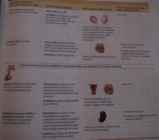
I stolled across this photo, which was very helpful to me when I was trying to understand the hypothalamus-pituitary complex and the hormones that this complex secretes. This photo shows the connections of the hypothalamus to the pituitary, and some of the targets of the pituitary hormones. It summerizes the hypothalamus- pituitary complex very well, and there wasn't alot of confusing extra information.


These charts on pages 530-531 in our textbook were a little more in depth in describing the hormones of the hypothalamus- pituitary complex and the effects of the hormones. This was very beneficial because it not only gave me the name of the hormones involved in this complex, but it gave me the regulation of release, target organs and effects, and the effects of hyposecretion and hypersecretion. So this chart provided me with more useful information, and the way it was organized helped me retain the information better.


Objective 34 & 37: Lymphatic and Immune system

Objective 34: Discuss the components and functions of the lymphatic and immune system
Objective 37: Compare the structure and function of the primary and secondary lymphatic organs and tissues

I found this diagram from Cornell University's website under the Biology section discussing the Lymphatic System. It is an awesome photo that summarizes the primary and secondary lymphatic organs and then summarizes the function or role that they play in the lymphatic and immune system. This picture was very helpful to me because the diagram only includes the components of the lymphatic and immune system, so it's not jumbled up with other veins, arteries, or organs in the body. When the chart is simple like this, it is easier for me to understand because it is overwhelming to see so much information at one time. I never really paid too much attention to how important the lymphatic and immune system was, since they are usually overlooked when thinking about major systems of the body. But without this important system, our cardiovascular system would stop working and our immune system would be very much impaired! When my mother was diagnosed with her rare disease Wegener's granulomatosis (that i mentioned on a previous slide), she had to take a lot of medication to be able to get herself in remission and her immune system was extremely damaged. I'm not exactly sure if it was the drugs she had to take, the disease itself, or maybe even both that caused the immune system to become almost useless. My mother could not be around anyone who was remotely sick, even anyone with a common cold, because she would catch the sickness ten times worse and they could result to pneumonia quick. I never thought too much about the lymphatic and immune system until my mother became sick, and now I have a better appreciation for what these two systems do for my body on a daily basis.


Objective 2 & 6: Thinking and communication skills

Objective 2: Apply critical and integrated thinking skills
Objective 6: Use written and oral communication skills to explain scientific concepts

This Anatomy and Physiology II class as a whole allowed me to use both these objectives every day. In the beginning of each lecture, our class had to complete recitation questions that allowed us to apply our critical and integrated thinking skills on the section that we were learning. We not only had to use the information from the section that we were learning in lecture, but we had to think back and remember information from previous sections and classes. Once we had the questions, we had to write answers down and discuss the questions with our classmates so we could figure out an answer and make sure that we understood the concept correctly. This method was very helpful because I had to use the textbook and use the information I learned from previous body systems and relate them to the question being asked. Being able to discuss with my class allowed me to use my oral communication skill, and also helped me understand some topics at time when I had no idea what the question was asking. Our lab time also assisted me with thinking and communication skills. I worked together and communicated with my group to understand and get our labs done. We discussed the lab questions together and relied on each other to figure out the answers. This e-portfolio is also another example of me having to use my thinking and communication skills. All of these objectives allow me to use my critical and integrated thinking skill and I am able to use my written communication when I report how I learned the information. I used oral skill in the process of this e-portfolio because I discussed with my classmates certain objectives that I was having trouble with and was able to get their input.

Monday, April 25, 2011

Objective 54: Address how erythropoietin regulates red blood cell production

The texbook does an amazing job at describing how erythropoietin regulates red blood cell production. It explains this process alot better than it does other, if I may add, because for some of the objectives I would have to search and search for something the soundeds somewhere near the answer. The process isn't extremely complicated and there aren't too many difficult words, which is why it was alot easier for me to comphend the first time I read it. The book also provides a great photo that summerizes the process, using a seesaw to demonstrate whether the body is in homeostasis or is imbalanced. The diagram shows the list of stimuluses that could distrub homeostasis in the body, and then shows step-by-step how the body reacts to bring itself back to normal homeostasis again. An example that helps me remember how important this process is to the body is athletes. Healthy athletes will inject EPO to provide them with increased stamina and performance, and will bring their normal hematocrit from 45% to almost as much as 65%. This could be deadly when the athlete dehydrates during a race, which will cause the blood to concentrate further and could cause the blood to because a thick, sticky "sludge". Then the athlete could have blood clotting, stroke, or even heart failure. With this example in mind and a very useful diagram, I am able to understand how important it is that erythropoietin regulates RBC production and how important it is not to tamper with our amazing body processes.


Objective 76: Differentiate between zygote, morula, and blastocyst

When I was trying to differentiate between zygote, morula and blastocyst I could not find anything about them in the textbook. I did, however, find information about the three of them in our lab book and it was very helpful. The lab book describes the accurately describes the three, and is straight to the point. The zygote is the fertilized egg, the morula is a solid ball of blastomeres, and the blastocyst consists of a sphere of trophoblast cells and a cell cluster called the inner cell mass. The diagram in the lab book made figuring out the differences of the three easier because I was able to see where each process took places within the uterus and also a picture of each cell structure. Being able to see where each process takes place in the uterus helps me understand the differences of the three better because I don't have to wonder by reading, I can actually see the pictures of the cell structures. This diagram refreshed my memory of information that I learned in my human growth and development class two years ago. I had just found out I was pregnant and was extremely scared and also a little bit curious on how the process of fertilizating really occurred. My teacher at the time was very animated and vocal, so when she came across this information she made it very interesting and with my new curiousity for the subject it kind of stuck. So this chart and clips of texts from the book were a great refresher.


Objective 3: Solve for unknowns by manipulating variables

This objective became clear to me when I had to do an experiment in lab that was testing for the presence of maltose. I actually can not locate the actual results that I had wrote down from the experiment, and we couldn't write on the papers that we had at our stations. But I had to do the experiment that determined how a solution reacted if there was a presence of maltose. This experiment allowed one of the test tubes to act as the control with just water and the Benedict's solution. The other test tube contained water, Benedict's solution, and a sugar solution. The two tubes were then placed in a beaker of boiling water for about 5-10 minutes. The control solution stayed the same, but the sugar solution tube began to change into a red-orange color which proved that there was a presence of maltose. This experiment helped me understand this objective because I had to manipulate the other test tube by adding the sugar solution to it, and then added that tube and the control tube into the beaker of boiling water. The boiling water manipulation is what helped me figure out the unknown information of whether the solution tested positive for maltose. I've completed numerious experiments in the past during my Chemisty course that involved having to manipulate variables to solve for unknown information. The manipulation was either adding a certain solution, placing tubes in boiling baths, using the bunsen burner, and the list could probably go on and on. This experiment reminded me of the experiments that I used to do in Chemisty. So this experiment was able to click that old lightbulb on.


Objective 31 & 32: Aorta and the Vena Cavas

Objective 31: Locate major arteries brancing off the aorta and regions of the body they supply blood to
Objective 32: Identify the major veins draining into the superior and inferior vena cavae


When I was trying to figure out these two objectives about the aorta and vena cavae, the end of chapter 18 was a HUGE help. It couldn't have been put any simpler. From pages 632-643, the textbook discussed the aorta and all the major arteries of the systemic circulation. This portion not only briefly discusses the major arteries, but it provides charts of where they all are in the body. The two charts that were most beneficial to me in understanding objetive 31 was the schematic flowchart on page 632 and the body's anterior view of the major arteries of the systemic circulation. Personally, I think the body's anterior view of all the major veins in the body was more helpful than the flow chart because I am able to see where certain veins are in relation to one another. I don't have to visualize where they may be located, I can actually see where they are and what portion of the body they reside in. The flowchart is still helpful though, because the diagram uses arrows to show where the arteries branch off at and share what part of body they supply blood to. On pages 644-652, the textbook starts to talk about the venae cavae and the major veins of the systemic circulation. This section also provides charts that show different regions of the body and the main veins that it houses. The pictures that aided me in understading the major veins draining into the superior and inferior vena cavae because it also provided not only the body's anterior view of the veins, but it also gave a schematic flowchart of the major veins. Along with these two charts, the book provides a description of the superior and inferior vena cavae and talks about the veins that drain into them. I overlooked this section at first because I didn't view it as important, but now i'm glad that it was included because it was beneficial. The visuals helped me see where the veins are in relation to the rest of the body and the flow chart helped me to see what veins actually drained into the vena cavaes.


Saturday, April 23, 2011

Objective 55: Trace the path of blood flow through the kidneys

This diagram found on page 840 of our textbook was very helpful in learning the path of the blood flow through the kidneys. It is a very simple diagram, but it isn't confusing and it is very easy to remember. I am able to learn the concept better if there happens to be a diagram included, instead of just reading the words from the text book so I was happy when I came across this. It shows the path of blood as journeys through one of the kidney's lobes and it continues the cycle until it gets to the last lobe. I also was able to locate a YouTube video of a girl showing and explaining the path of the blood flow through the kidneys on a model in her class. This is helpful because I not only can read the chart, but I can hear someone explaining it to me while showing me on a model. This helped my visual learning. For some reason I thought that the blood flow through the kidneys would have been a more complicated one, but I'm relieved to find out that it is relatively simple. That helps me out tremendously.


Objective 62: Describe external and internal gross renal system anatomy

When I was trying to learn the external and internal anatomy of the renal system, I mainly referred to my textbook and slide outlines because they provided me with many visual diagrams. The slide outlines for the chapter 24 provided some basic information that described the different characteristics and internal layers of the organs. Since I am a visual learner, the diagrams were amazing in helping me understand the placement of the organs in the body. Our lab dissection was where the light bulb really clicked on for me though. Dissection 8 was the Urinary System of the Cat and I was able to see first hand how the organs were actually connected and place in relation to one another. Its one thing to see it in the diagram, but it seemed to make a whole lot more sense when I was actually dissecting the cat and seeing the organs for myself. This dissection instructed me to dissect into the kidney (the first picture is the actual kidney I dissected) and the bladder. I'm a hands-on person as well, so being able to see the cortex and medulla of the kidney and also the internal sphincter of the bladder really helped me remember them. I kind of dreaded the dissections at first because I was also afraid that I was going to cut the cat wrong, but I am now glad that I had the opportunity to do the dissection in lab.