Tuesday, April 26, 2011

Objective 5 & 12: Hypothalamus and hypothalamus- pituitary complex

Objective 5: Explain why the hypothalamus is classified as an endocrine gland
Objective 12: Name hypothalamus- pituitary complex hormones and give their functions

Is the hypothalamus an endocrine gland? It seems to be an ongoing debate even to this day. A true endocrine gland is a ductless gland that releases hormones into surrounding tissue fluid and has a rich vascular and lymphatic drainage to recieve the hormones. But the hypothalamus is an amazing gland. It not only performs neural functions and controls the endocrine system, but it also produces and releases hormones. Producing and releasing hormones is the major factor in deciding whether or not the gland is an endocrine gland, and the hypothalamus has that. I agree when it is said that the hypothalamus gland is an endocrine gland.

I stolled across this photo, which was very helpful to me when I was trying to understand the hypothalamus-pituitary complex and the hormones that this complex secretes. This photo shows the connections of the hypothalamus to the pituitary, and some of the targets of the pituitary hormones. It summerizes the hypothalamus- pituitary complex very well, and there wasn't alot of confusing extra information.


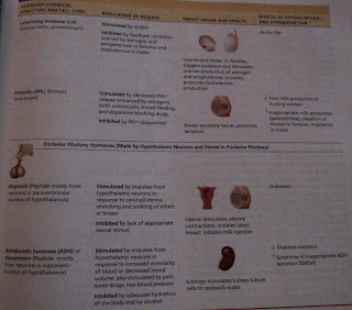
These charts on pages 530-531 in our textbook were a little more in depth in describing the hormones of the hypothalamus- pituitary complex and the effects of the hormones. This was very beneficial because it not only gave me the name of the hormones involved in this complex, but it gave me the regulation of release, target organs and effects, and the effects of hyposecretion and hypersecretion. So this chart provided me with more useful information, and the way it was organized helped me retain the information better.


Objective 34 & 37: Lymphatic and Immune system

Objective 34: Discuss the components and functions of the lymphatic and immune system
Objective 37: Compare the structure and function of the primary and secondary lymphatic organs and tissues

I found this diagram from Cornell University's website under the Biology section discussing the Lymphatic System. It is an awesome photo that summarizes the primary and secondary lymphatic organs and then summarizes the function or role that they play in the lymphatic and immune system. This picture was very helpful to me because the diagram only includes the components of the lymphatic and immune system, so it's not jumbled up with other veins, arteries, or organs in the body. When the chart is simple like this, it is easier for me to understand because it is overwhelming to see so much information at one time. I never really paid too much attention to how important the lymphatic and immune system was, since they are usually overlooked when thinking about major systems of the body. But without this important system, our cardiovascular system would stop working and our immune system would be very much impaired! When my mother was diagnosed with her rare disease Wegener's granulomatosis (that i mentioned on a previous slide), she had to take a lot of medication to be able to get herself in remission and her immune system was extremely damaged. I'm not exactly sure if it was the drugs she had to take, the disease itself, or maybe even both that caused the immune system to become almost useless. My mother could not be around anyone who was remotely sick, even anyone with a common cold, because she would catch the sickness ten times worse and they could result to pneumonia quick. I never thought too much about the lymphatic and immune system until my mother became sick, and now I have a better appreciation for what these two systems do for my body on a daily basis.


Objective 2 & 6: Thinking and communication skills

Objective 2: Apply critical and integrated thinking skills
Objective 6: Use written and oral communication skills to explain scientific concepts

This Anatomy and Physiology II class as a whole allowed me to use both these objectives every day. In the beginning of each lecture, our class had to complete recitation questions that allowed us to apply our critical and integrated thinking skills on the section that we were learning. We not only had to use the information from the section that we were learning in lecture, but we had to think back and remember information from previous sections and classes. Once we had the questions, we had to write answers down and discuss the questions with our classmates so we could figure out an answer and make sure that we understood the concept correctly. This method was very helpful because I had to use the textbook and use the information I learned from previous body systems and relate them to the question being asked. Being able to discuss with my class allowed me to use my oral communication skill, and also helped me understand some topics at time when I had no idea what the question was asking. Our lab time also assisted me with thinking and communication skills. I worked together and communicated with my group to understand and get our labs done. We discussed the lab questions together and relied on each other to figure out the answers. This e-portfolio is also another example of me having to use my thinking and communication skills. All of these objectives allow me to use my critical and integrated thinking skill and I am able to use my written communication when I report how I learned the information. I used oral skill in the process of this e-portfolio because I discussed with my classmates certain objectives that I was having trouble with and was able to get their input.

Monday, April 25, 2011

Objective 54: Address how erythropoietin regulates red blood cell production

The texbook does an amazing job at describing how erythropoietin regulates red blood cell production. It explains this process alot better than it does other, if I may add, because for some of the objectives I would have to search and search for something the soundeds somewhere near the answer. The process isn't extremely complicated and there aren't too many difficult words, which is why it was alot easier for me to comphend the first time I read it. The book also provides a great photo that summerizes the process, using a seesaw to demonstrate whether the body is in homeostasis or is imbalanced. The diagram shows the list of stimuluses that could distrub homeostasis in the body, and then shows step-by-step how the body reacts to bring itself back to normal homeostasis again. An example that helps me remember how important this process is to the body is athletes. Healthy athletes will inject EPO to provide them with increased stamina and performance, and will bring their normal hematocrit from 45% to almost as much as 65%. This could be deadly when the athlete dehydrates during a race, which will cause the blood to concentrate further and could cause the blood to because a thick, sticky "sludge". Then the athlete could have blood clotting, stroke, or even heart failure. With this example in mind and a very useful diagram, I am able to understand how important it is that erythropoietin regulates RBC production and how important it is not to tamper with our amazing body processes.


Objective 76: Differentiate between zygote, morula, and blastocyst

When I was trying to differentiate between zygote, morula and blastocyst I could not find anything about them in the textbook. I did, however, find information about the three of them in our lab book and it was very helpful. The lab book describes the accurately describes the three, and is straight to the point. The zygote is the fertilized egg, the morula is a solid ball of blastomeres, and the blastocyst consists of a sphere of trophoblast cells and a cell cluster called the inner cell mass. The diagram in the lab book made figuring out the differences of the three easier because I was able to see where each process took places within the uterus and also a picture of each cell structure. Being able to see where each process takes place in the uterus helps me understand the differences of the three better because I don't have to wonder by reading, I can actually see the pictures of the cell structures. This diagram refreshed my memory of information that I learned in my human growth and development class two years ago. I had just found out I was pregnant and was extremely scared and also a little bit curious on how the process of fertilizating really occurred. My teacher at the time was very animated and vocal, so when she came across this information she made it very interesting and with my new curiousity for the subject it kind of stuck. So this chart and clips of texts from the book were a great refresher.


Objective 3: Solve for unknowns by manipulating variables

This objective became clear to me when I had to do an experiment in lab that was testing for the presence of maltose. I actually can not locate the actual results that I had wrote down from the experiment, and we couldn't write on the papers that we had at our stations. But I had to do the experiment that determined how a solution reacted if there was a presence of maltose. This experiment allowed one of the test tubes to act as the control with just water and the Benedict's solution. The other test tube contained water, Benedict's solution, and a sugar solution. The two tubes were then placed in a beaker of boiling water for about 5-10 minutes. The control solution stayed the same, but the sugar solution tube began to change into a red-orange color which proved that there was a presence of maltose. This experiment helped me understand this objective because I had to manipulate the other test tube by adding the sugar solution to it, and then added that tube and the control tube into the beaker of boiling water. The boiling water manipulation is what helped me figure out the unknown information of whether the solution tested positive for maltose. I've completed numerious experiments in the past during my Chemisty course that involved having to manipulate variables to solve for unknown information. The manipulation was either adding a certain solution, placing tubes in boiling baths, using the bunsen burner, and the list could probably go on and on. This experiment reminded me of the experiments that I used to do in Chemisty. So this experiment was able to click that old lightbulb on.


Objective 31 & 32: Aorta and the Vena Cavas

Objective 31: Locate major arteries brancing off the aorta and regions of the body they supply blood to
Objective 32: Identify the major veins draining into the superior and inferior vena cavae


When I was trying to figure out these two objectives about the aorta and vena cavae, the end of chapter 18 was a HUGE help. It couldn't have been put any simpler. From pages 632-643, the textbook discussed the aorta and all the major arteries of the systemic circulation. This portion not only briefly discusses the major arteries, but it provides charts of where they all are in the body. The two charts that were most beneficial to me in understanding objetive 31 was the schematic flowchart on page 632 and the body's anterior view of the major arteries of the systemic circulation. Personally, I think the body's anterior view of all the major veins in the body was more helpful than the flow chart because I am able to see where certain veins are in relation to one another. I don't have to visualize where they may be located, I can actually see where they are and what portion of the body they reside in. The flowchart is still helpful though, because the diagram uses arrows to show where the arteries branch off at and share what part of body they supply blood to. On pages 644-652, the textbook starts to talk about the venae cavae and the major veins of the systemic circulation. This section also provides charts that show different regions of the body and the main veins that it houses. The pictures that aided me in understading the major veins draining into the superior and inferior vena cavae because it also provided not only the body's anterior view of the veins, but it also gave a schematic flowchart of the major veins. Along with these two charts, the book provides a description of the superior and inferior vena cavae and talks about the veins that drain into them. I overlooked this section at first because I didn't view it as important, but now i'm glad that it was included because it was beneficial. The visuals helped me see where the veins are in relation to the rest of the body and the flow chart helped me to see what veins actually drained into the vena cavaes.